38 adipose tissue with labels
Solved Drag the labels onto the diagram to identify the - Chegg Anatomy and Physiology questions and answers. Drag the labels onto the diagram to identify the parts of the kidney. Reset Help Minor calyx Hilum Ureter Adipose tissue in renal sinus Renal pelvis Renal sinus Major calyx Connection to minor calyx. Adipose Tissue - Composition, Location and Function Adipose, or fat, tissue is loose connective tissue composed of fat cells known as adipocytes. Adipocytes contain lipid droplets of stored triglycerides. These cells swell as they store fat and shrink when the fat is used for energy. Adipose tissue helps to store energy in the form of fat, cushion internal organs, and insulate the body.
Adipose connective tissue - University of Wisconsin-La Crosse Adipose connective tissue 1. Cell membrane 2. Cell nucleus 3. Fat vacuoles Adipose connective tissue cells are specialized for fat storage and do not form ground substance or fibers. On prepared slides, adipose tissue appears somewhat like a fish net with white spaces connected together in a network.

Adipose tissue with labels
A&P Flashcards | Quizlet Classify the body location/structure with the correct type of connective tissue by clicking and dragging the labels from column A to the appropriate location in column B. adipose -subcutaneous layer-yellow bone marrow elastic connective-vocal cords-larger artery walls compact bone -bone shafts-spongy bone-ends of long bones -reticular connective -red bone … Off-label use of adipose-derived stem cells - ScienceDirect 1. Introduction. Recent advances in regenerative medicine, particularly the discovery of multipotent, easily accessible stem cells such as adipose-derived stem cells (ASCs), have provided the opportunity to use autologous stem cell transplants as regenerative therapies .Fat is an active and dynamic tissue composed of several different cell types, including adipocytes, fibroblasts, smooth ... AKT antibody (60203-2-Ig) | Proteintech mouse brain tissue were subjected to SDS PAGE followed by western blot with 60203-2-Ig (AKT Antibody) at dilution of 1:1000 incubated at room temperature for 1.5 hours. IHC Figures IHC staining of human breast cancer using 60203-2-Ig
Adipose tissue with labels. Brown Adipocytes - Yale University Brown adipose tissue is typically found in large amounts in newborns and some hibernating animals and is important as a source of energy. The cells in brown fat contain numerous and very distinct lipid droplets. The presence of an uncoupling protein in these cells causes the generation of heat that allows for non-shivering thermogenesis. Two-photon excited fluorescence of intrinsic fluorophores enables label ... Adipose tissue function has been recognized to exert significant influence on systemic metabolic balance and overall homeostasis health through energy storage via lipid accumulation, direct energy expenditure through substrate oxidation 1,2, and secretion of various signaling and regulatory molecules 3,4.Major health problems such as type 2 diabetes mellitus, cancer, and cardiovascular disease ... SC 2115 Anatomy and Physiology I - Middlesex Community College Draw and label Reticular Tissue: reticular fibers form the stroma . E. Adipose Tissue: surrounds heart and kidneys, subcutaneous tissue, and greater omentum. This is the most easily recognized tissue and will be found widely distributed in every organ microscopically studied this year. Lab Notes Home Page - Kentucky Community and Technical College System Types of Connective Tissue. A). Connective Tissue Proper. 1). Loose Connective Tissue. i). Areolar Connective Tissue. Matrix with fibroblasts supported by gel membrane with collagenous & elastic fibers. ii). Adipose Tissue (fat) Tightly packed & highly vascularized An oil droplet pushes the nucleus to one side. iii).
The developmental origins of adipose tissue Adipose tissue is formed at stereotypic times and locations in a diverse array of organisms. Once formed, the tissue is dynamic, responding to homeostatic and external cues and capable of a 15-fold expansion. The formation and maintenance of adipose tissue is essential to many biological processes and when perturbed leads to significant diseases. Adipose tissue - Vida Private Label Adipose tissue. Is it possible to act on adipose tissue, and how? A treatment aimed at reducing fat deposits and localised oedemas. Fat deposits. Adipose tissue: a fat lot of good? | Society for Endocrinology 3D rendering of a 100-µm thick section of murine adipose tissue in the kidney. Immunofluorescent staining for the lipid droplet protein perilipin is in cyan. A lineage tracing Cre-induced tdTomato fluorescent protein labels a subset of adipocytes red. In the centre, the nucleus of a resident adipose stem cell is labelled green. ©J. Rochford Histology - Yale University The gross tissue structure of the thymus depends upon the age of the individual. The organ is large in early life and filled with lymphocytes, but involutes with advancing age, as the parenchyma is gradually replaced by adipose tissue.
Brown Adipose Tissue | Connective Tissue Brown Adipocytes. Brown fat is specialized for the generation of heat (thermogenesis). In adult humans, it is found in small amounts in the upper chest and neck. Brown adipocytes are smaller cells with a centrally located nucleus and many small lipid droplets (multilocular). The abundance of mitochondria accounts for the brown color characteristic of its gross appearance. Adipose connective tissue - Austin Community College District Adipose connective tissue 400X The bar labeled "a" indicates the width of one adipose cell (adipocyte). The light purple dots you see inside the cells are an artifact of process used to make the images, and do not represent real structures. The arrow points to the nucleus of one adipocyte. Adipose tissue: Definition, location, function | Kenhub Brown adipose tissue labeled (histological slide) In contrast to white adipocytes, brown adipocytes have the appearance of a sponge due to the multiple droplets in the cytoplasm. Groups of adipocytes are divided into lobules by connective septa, which contain a substantial amount of blood vessels and unmyelinated nerve fibers. Solved Label the components of adipose tissue. Fibroblast | Chegg.com Label the components of adipose tissue. Fibroblast Cell membrane Elastic fiber Nucleus Adipocyte ; Question: Label the components of adipose tissue. Fibroblast Cell membrane Elastic fiber Nucleus Adipocyte

Quia - PRENATAL - FEMALE REPRODUCTIVE LABELS
The Genotype-Tissue Expression (GTEx) pilot analysis: Multitissue … 08.05.2015 · Study design. During the pilot, we recruited 237 postmortem donors, collecting an average of 28 tissue samples per donor spanning 54 distinct body sites (fig. S1 and tables S1 and S2).Blood-derived DNA samples were genotyped at approximately 4.3 million sites, with additional variants imputed using the 1000 Genomes phase I, resulting in ~6.8 million single-nucleotide …
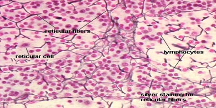
Reticular Connective Tissue - Assignment Point
Adipose tissue (labels) - histology slide Histology Photo Album Home Album list Last uploads Last comments Most viewed Top rated My Favorites Search FILE 23/27 Adipose tissue (labels) - histology slide This slide shows hyaline cartilage, adipose tissue, and skeletal muscle. Histology slide courtesy of William L. Todt, Ph.D. at Concordia College, Moorhead, Minnesota.
[Solved] V Part3 Complete the Part B Concept Map to indicate connective tissues' charactelistic ...
Origin and Functions of Tissue Macrophages - PMC 17.07.2014 · labels monocytes, which allows for differential targeting: is limited to tissue macrophages that express CX3CR1 at the time of induction: Yona et al. (2013); Goldmann et al. (2013); Parkhurst et al. (2013) Rag1 Cre: identifies lymphomyeloid progenitors: is the only technique for specifically labeling lymphomyeloid progenitors: it is unclear whether progeny …
Post a Comment for "38 adipose tissue with labels"